Eyelid Lesion Removal
An eyelid lesion is a pathological change in the tissue of the eyelid. There are many types of lesions, most of which are benign or harmless. However, some lesions may be malignant or cancerous and require careful biopsy and sometimes referral to a dermatologic specialist.
An eyelid lesion may be pigmented or colored. These range from relatively common disorders such as styes or chalazia, which are small red bumps on our eyelids to other types of benign eyelid lesions including seborrheic keratoses, papillomas, nevi, hemangiomas, actinic keratosis and xanthelasmas.
The 3 most common types of cancerous lesions of the eyelid are basal cell carcinomas (BCC), Squamous cell carcinomas (SCC) and malignant melanomas. If your ophthalmologist suspects a lesion could be cancerous he may recommend excisional biopsy. If a cancer is confirmed by biopsy it usually results in a referral to dermatologic specialist familiar with Mohs surgery which is a tissue sparing procedure that can more completely remove the cancer while sparing as much eyelid/facial tissue as possible.
How Are Eyelid Lesions Detected?
How Are Eyelid Lesions Treated?
In most cases, benign eyelid lesions require no treatment. However, the lesion can be surgically removed from the eyelid for cosmetic reasons, if it interferes with your vision, or if your doctor sees anything abnormal. Any lesion that is surgically removed will normally be sent for biopsy.
During a surgical excision of an eyelid lesion, you will feel little or no discomfort. On the day of treatment, a staff member will welcome you. The staff will help you prepare for your surgery by putting eye drops in your eye.
An anesthetic injection is given around the eye to prevent discomfort during the removal process. A driver may be recommended. The procedure normally takes less than 30 minutes. Typically, no eye patch is needed after the surgery.
Your eye may appear to be slightly bruised and swollen following the surgery. However, this should not cause significant discomfort. Your surgeon will follow your progress to monitor for signs of inflammation and infection.
Some potential complications from surgery include the risk of bleeding, infection and hematoma, which can usually be successfully treated.
Common Benign Eyelid Lesions
Seborrheic Keratoses
Seborrheic keratoses (SK) are a common type of benign tumor and are associated with aging. SKs can occur anywhere on the body, including the eyelids, and can range in color from light tan to dark brown or black. Once you have one SK, you are likely to develop more of them. These growths, which look like they’re stuck onto the surface of your skin, are usually uneven in texture, and can be very itchy. Scratching them can cause bleeding and can also lead to a skin infection. Normally treatment of SKs is not necessary. However, an SK can be surgically removed from the eyelid for cosmetic reasons, if it interferes with your vision, or if your doctor sees anything abnormal. When an SK is removed, the tissue is usually sent for a biopsy for precautionary reasons.
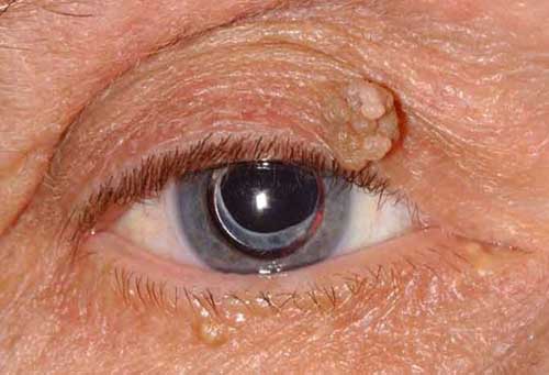
Papilloma
A papilloma is smooth, round benign tumor that originates in the outer layer of skin on your eyelid. It is also called a squamous papilloma. This type of lesion is a common type of benign eyelid tumor and is associated with aging. This type of papilloma is commonly called a “skin tag” when located on other parts of your body.
In some cases, a papilloma can be caused by a viral infection and are known as “viral warts”. Viral papillomas are more common in children and teenagers. They can have a fleshy, lobular appearance like cauliflower. They also can continue to grow larger.
Eyelid papillomas rarely require removal for medical reasons. They can easily be removed in an outpatient procedure if necessary for cosmetic purposes of if they interfere with vision.
Nevi or Nevus
A nevus is a benign neoplasm made up of overgrowth of melanocytes. In other words, it can be described as a mole. A nevus can be present at birth but may also develop after birth, usually appearing in parts of the body exposed to sunlight. Normally moles on your eyelids would not be removed for medical reasons unless they interfere with your vision. However, if you notice changes in the shape of your moles, if they become painful, itchy or bleed, they should be removed and subjected to a biopsy to rule out melanomas.
Hemangioama
Hemangioma is a benign tumor of the endothelial cells that line blood vessels. This type of tumor appears shortly after birth or in infancy and grows rapidly for an initial period of time. Then, the tumor will gradually become smaller and usually resolve itself by the time the child reaches age 7. However, if this growth is situated near your child’s eye, it can have a serious impact on his or her vision and it is highly recommended that you seek out an ophthalmologist to evaluate it.
Actinic Keratosis
Actinic keratosis (AK) is a scaly white patch that forms on the surface of your skin, usually in areas exposed to sunlight. Although AKs are benign, they are also considered to be pre-cancerous lesions. If you have an AK on your eyelid, our surgeons can remove it and send a sample of the t issue to a pathologist for biopsy. If cancer is found a recommendation to a dermatologic specialist for a more complete resection may be deemed necessary.
Xanthelasma
Xanthelasma are lesions of yellow plaque on the eyelids. They can be soft, semi-solid or hard and have a tendency to spread and to become permanent. They occur frequently in people with elevated cholesterol levels and / or reduced levels of HDL, the “good” cholesterol. Xanthelasma can also be associated with uncontrolled diabetes. Although these lesions are benign, they may be unsightly and patients often want them removed for cosmetic reasons. If you have xanthelasma, the usual treatment is surgical removal, which can be performed in the office. There is a small chance of recurrence, especially in those with cholesterol abnormality.
Experience You Can See
Thibodaux : 985-446-0506 | Houma : 985-879-2393